פרופ' הרצל בן-חור: קורות חיים ופרסומים
בס"ד
CURRICULUM VITAE- Professor Herzel Ben Hur
FACULTY/DEPT OBSTETRICS & Gynecology, Gynecological Oncology
HOSPITAL/DEPT Assaf Harofeh Medical Center
Email: herzelb@asaf.health.gov.il, herzelb1@gmail.com
PLACE OF BIRTH IRAN
DATE OF ARRIVAL IN ISRAEL 1957
ZAHAL (ISRAELI) MILITARY SERVICE 1967- 1970. Still serving in reserves Rank Colonel
Status: Married, children 4
Degree: M.D CV: 3rd of August 2009
A. Education
PERIODS OF STUDIES (DATES) 1970-72: University of Siena Italy
1973-77: Sackler School of Medicine, Tel Aviv University Israel. M.D.
26.06.1978
(Date Awarded)
Title of Doctoral Dissertation
Names of Supervisors Alterations of the colonic flora and their effect on the hydrogen breath test.
Gilat T, Ben Hur H Gelman Malachi E, Terdiman R, PeledY.
Gut; 19(7), 602-5, 1978. (Ref 1)
B. FURTHER STUDIES
Period of Study (dates) 1978-1984 Residency in Obstetrics and Gynecology, Kaplan Hospital, Rehovot (Affiliated to the Hebrew University Medical School, Jersalem, Israel).
1984 Board certification in obstetrics and gynecology
Specialist of Obstetrics & Gynecology
No: 08383
29.03.1985
Date Awarded
C. ACADEMIC AND PROFESSIONAL EXPERIENCE
(FOR MDs – ACADEMIC EXPERIENCE; CLINICAL EXPERIENCE)
Period (Dates) Advanced Surgical Training
1985 Postgraduate training in Gyneco-Oncological Surgery, Queen Elizabeth Hospital, Gateshead, Newcastle, England (Mr. J. M. Monaghan).
1987 Postgraduate training in General and Vascular Surgery, Kaplan Hospital, Rehovot (Chairmen: Prof. R. Pffeferman and Dr. A. Mashiah).
1987-1994 Several two months courses of training and refinement of surgical skills, Gateshead England.
1997 Residential course (30 d) in Advanced Gynecological Laparoscopic (Level 4) and Hysteroscopic (Level 3) Surgery. Mr Dr. J. H. Phipps, Nuneaton, UK.
1997-1998 Several one week Residential courses of Vaginal Surgery at Edouard Heriot Hospital, Mr. D. Dargeant Lyon France.
Academic and Professional Responsibilities
1982 Instructor, Hebrew University School of Meine Jerusalem.
1982-1995 In charge of fifth year medical students clerkship in obstetrics and gynecology, Kaplan Hospital Rehovot.
1986 Consultant, Obstetrics & Gynecology, Kaplan Hospital, Rehovot
1991 – 2001 Deputy Head, Department of Obstetrics and Gynecology, Kaplan Medical Center, Rehovot Israel.
1993 – Present Visiting Scientist, Weizmann Institute of Science, Rehovot.
1996 – 2001 Senior Lecturer, School of Medicine, Hebrew University Jerusalem
1997- 2001 Mentoring and supervision of MD dissertations.
Kaplan Medical Center, Rehovot affiliated to the Hebrew University Jerusalem
1997- 2001 Mentoring and supervision of residents. Advanced training and research in Basic Sciences.
2001- Present, Senior Obstetrician, Gynecologist & Gyneco-Oncologist, General Health Services, Central District, Lod Center, Israel.
2001 – Present, Senior Obstetrician, Gynecologist & Gyneco-Oncologist, Assaf Harofeh Medical Center.
2002 – Present, Head, Laboratory for Early Detection,Weizmann Park, Ness Ziona, Israel.
D. ACTIVE PARTICIPATION IN SCIENTIFIC MEETINGS
Please see Abstracts below, 59 Abstracts.
E. ACADEMIC AND PROFESSIONAL AWARDS
1982 Recipient of Rogozinsky Prize for outstanding work as Senior Registrar.
1999 Selected by students in the Faculty of Medicine among the "Best Teachers" in the Hebrew University Medical School, Jerusalem.
Grants
1993 From the Chief Scientist of the Israeli Ministry of Health, for Expression of Estrogen & Progesterone receptors in normal and pathological human veins.
(Prizes, fellowships, grants, scholarships, etc)
F. MEMBERSHIP IN PROFESSIONAL SOCIETIES
Year 1979 Israel Medical Association
1984 Israel Society of Obstetrics & Gynecology
1987 Israel Society of Gynecological Oncology
1988 Israel Society of Fertility & Sterility
l994 European Society of Gynaecologic Oncology
2000 Israel Society of Colposcopy and Cervical Pathology
G. DOCTORAL STUDENTS SUPERVISED BY CANDIDATE
Training and instruction of students and residents:
MD Students
1. A. Ben- Meir
MD dissertation. Hebrew University Medical School, Jerusalem. 1998.
Publications:
a. Tumor-suppressor effects of vaccination with the soluble p53 antigen-bearing liposomes on chemically induced skin cancer in mice. Thesis of MD dissertation. Hebrew University Medical School, Jerusalem, 1998.
Anticancer Res; 18(6A), 4237-42, 1998, (Ref 47).
2. M. Ashkenazi
MD dissertation. Hebrew University Medical School, Jerusalem.1999.
The relationship between the splenic lymphoid system of a
host and implanted hepatoma cells.
Publications:
a. Lymphoid elements and apoptosis-related proteins (Fas, Fas ligand, p53 and bcl-2) in lichen sclerosus and carcinoma of the vulva in human.
J Gynecol Oncol; 22, 104-109, 2001, (Ref 75).
Basic science
1. E. Plonsky – Basic Sciences Research, Department of Urology, Kaplan Medical Center, Rehovot, 1999.
Publications:
a. Morphological and morphometric studies of the splenic antitumor immune response, elicited by liposome-covered soluble p53 kDa antigen, in chemically-induced rat colon cancer.
Int J Mol Medicine; 3(5), 545-9, 1999, (Ref 52).
b.E. Plonski – Transplacental effects of IgG generated against soluble 53 kDa protein on the splenic lymph system of rat progeny exposed to carcinogen: rate of apoptosis, proliferation of lymphocytes and expression of Fas and Fas ligand proteins. Eur J Gynaecol Oncol; 20 (4), 306-310, 1999, (Ref 55).
2. A. Budovski – Basic Sciences Research, Department of
Anesthesiology, Kaplan Medical Center, Rehovot, 2000.
Publications:
a. Transplacental effects of maternal feeding with high fat diets on
the splenic lymphatic system in offspring exposed to a low dose of carcinogen.
Int J Molec Med; 6(3), 337-431, 2000, (Ref 70).
3. O. Cohen – Basic Sciences Research, Department of Obstet.& Gynocol., Assaf Harofeh Medical Center, Tzerifin, 2001.
Publications:
a. Transplacental effect of a 15% olive-oil diet on functional
activity of immune components in the spleen and colon tumors
of rat offspring. Oncol Rep; 8, 1045-1049, 2001, (Ref 82).
b. The role of lymphocytes and macrophages in human breast
tumorigenesis: An immunohistochemical and morphometric
study. Anticancer Res; 22, 1231-1238, 2002, (Ref 89).
4. E. Mordechai – Basic Sciences Research, Department of Obstet. & Gynocol., Assaf Harofeh Medical Center, Tzerifin, 2002.
Publications:
a. Apoptosis-related proteins (Bcl-2, p53, Fas and Fas ligand) in different types of human breast tumors. Oncol Rep; 9, 977-980, 2002, (Ref 96).
PhD Students:
1. Ronit Abir–Hadassa School of Medicine, Jerusalem
a. The effects of serum from women with miscarriages on the in vitro development of mouse preimplantation embryos.
Acta Obstet Gynecol Scand 1990; 69(1):27-33, (Ref 10).
b. Correlation between yolk sac damage and embryonic
anomalies induced by serum from women with recurrent
abortions in early somite rat embryos in culture.
Teratology 1992; 45(5):l62, 502-503, l992, (Ref 17).
c. IgG exchange as a mean of partial correction of malformations
induced by recurrent abortions sera to rat embryos in vitro.
Toxicol In Vitro 1993; 7:8l7-826, (Ref 20).
2. Gil Mor–Weizmann Institute of Science, Rehovot
a. Evidence for the presence of estrogen receptors in monocytes. Endocr J 1993; l:387-395, (Ref 22) .
b. Menopause is associated with a significant increase in blood monocyte number and a relative decrease in the expression of estrogen receptors in human peripheral monocytes. Am J Reprod Immunol 1995; 34(6):363-9, (Ref 21).
c. Menopause is associated with a significant increase in blood monocyte number and a relative decrease in the expression of estrogen receptors in human peripheral monocytes. Am J Reprod Immunol 1995; 34(6):363-9, (Ref 33).
3 . Giora Alon, The Faculty of Agriculture, Rehovot
Small intestine development in the young chick: Crypt formation and enterocyte proliferation and migration. British Poultry Science. 2000, 41:544-551, (Ref 72).
4 . Orna Bar Peled – Weizmann Institute of Science Rehovot.
a. Fetal human brain exhibits a prenatal peak in the density
of serotonin 5-HT1A receptors.
Neurosci Lett 1991; 127(2):173-6, (Ref 12).
c. Developmental pattern of muscarinic receptors in normal and Down's syndrome fetal brain–an autoradiographic study. Neurosci Lett; 133(2), 154-8, 1991. (Ref 14).
d. Distribution of glutamate transporter subtypes during human brain development. J Neurochem 1997; 69(6):2571-80, (Ref 41).
RESEARCH ACCOMPLISHMENTS
Research activities to date
From 1984 to date I have been involved in the following areas of research:
I. Embryonic development (1984-1997)
II. Detection of steroid receptors in reproductive and non-reproductive systems (1990-1995)
III. Development and detection of cancers (1995- to date)
IV. The role of immune reaction in cancer development and suppression (1984 to date).
V. Secretory immune system (sis), in embryonic and fetal, normal and pathological tissues (2000-to date).
VI. Collaborative research with the scientists in various institutions, primarily with scientists from the Weizmann Institute of Science (1984- to date).
Below is a brief summary of my research activities in the above fields:
Embryonic development
Early abortion and embryonic malformations were the focus of several of my studies. (Ref: 7 10 12 17 20 23 29 36 41).
Main conclusion from these studies: Sera from women that had suffered recurrent miscarriages could increase the rate of abortion in experimental animals.
Detection of steroid receptors in reproductive and non reproductive systems
(Ref: 21 22 32 33 39 50).
General considerations
Estrogens affect many systems in the body such as the reproductive, immune, skeletal systems. It can be expected that the immunoregulatory properties of estrogens are possibly mediated by binding of the hormone to the intracellular receptors present in target cells (e.g.monocytes/macrophages, see Figure 1).
 Figure 1: A model of endocrine-immune modulation involving the monocyte/macrophage system. Estrogen synthesized and secreted by the
ovarian cells immunomodulate its effects via the estrogen receptor (ER) present in the monocytes.
Interestingly, in 1990 there was no definite evidence of the presence of ER in the cells of the immune system and reagents (e.g. specific antibodies to steroid receptors) were not commercially available for the determination of ER in non reproductive systems (immune, bone) and reproductive system (endometrium). Accordingly, for the detection of ER in these systems we used a novel reagent, an anti-idiotypic antibody to anti-estradiol, clone 1D5 that interacts with the estradiol receptor (ER). For this purpose we investigated the expression of ER
Name: Herzel Ben Hur Degree: M.D CV: 3rd of August 2009
in relation to sub-populations of human peripheral blood mononuclear cells (PBMC) using fluorescence activated cell sorter (FACS). Dual color flow cytometry of PMBC indicated that a dimly labeled CD4 positive population was stained with anti-CD14, a specific marker for monocytes as well as with anti-idiotypic antibody clone 1D5 that interacts with ER. (33).
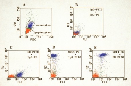
Figure 2. FACS analysis of the expression of CD14+ (monocyte marker) and estrogen receptor cell population by direct staining of PBMC with a mixture of anti-CD14 PE and FITC conjugate of anti-idiotypic antibody 1D5 which recognizes ER. Note that 88% of anti-CD14+ cells are positive to ER.
Using this method we evaluated the clinical significance of the differential expression of estrogen receptor (ER) in human monocytes. Conclusions: During menopause there is a significant decrease in the percentage of ER positive monocytes, and an increase in blood monocyte number, which declines following estrogen replacement therapy to values of the young. These findings suggest that estrogen modulates the monocyte numbers and its effects may be mediated through the ER in the monocytes (33).
Human endometrium
In the normal human endometrium, the intensity of ER staining varied according to the phase of the cycle as well as according to the cell type. On the other hand, endometrial ER evaluation of the patient with DES syndrome showed minimal expression of ER and after treatment with conjugated estrogens, endometrial biopsy revealed a significant increase in ER expression.
Conclusions: These findings suggest that changes in ER expression can be monitored in the endometrium during hormonal therapy (32) Human cartilage.
Our work also demonstrated their intranuclear and perinuclear localization by immunostaining, immunofluorescence and autoradiography in fetal cartilage.
And we can now analyze and quantify various hormone receptors in many different tissues of interest by immunocytochemistry and image analysis.
Development and detection of cancer
(Ref: 28 34 35 37 38 46 53-55 61 64 67 68 70 71 73-78 87 89 95-98 101 111-115 124 127-129 132).
The roles of apoptosis, apoptosis related proteins (Fas, FasL, bcl-2 and p53), and their interaction with lymphoid immune elements have been studied in cancers of the colon, endometrium, ovaries, skin, the mammary gland, and the prostate. Different components of the immune system become active during different stages of tumor development. Moderate reaction of T killer cells and macrophages is typical in benign cysts. In borderline tumors, the activity of killer cells increases in the parenchyma, and that of T helper cells and of macrophages in the stroma. In carcinomas with high lymphoid infiltration, a strong macrophagic reaction in the tumoral parenchyma is typical.
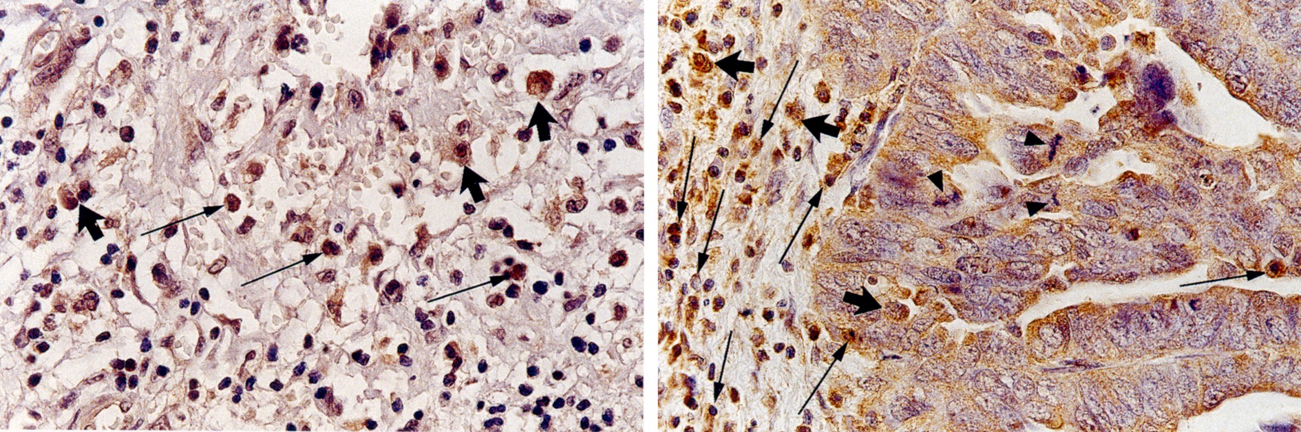
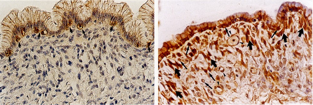
Figure 3: Serous cystoma and mucous borderline tumor: expression of Fas and bcl-2, and Fas-ligand (Ref 64).
Soluble p53 and Fas are effective markers of gynecological cancers that could be used for early diagnosis of malignant and pre-cancerous states. We showed a possibility to use the determination of serum-levels of p53 antigen for detection colorectal and endometrial cancer. Vaccination with soluble p53 antigen was shown to be tumor-suppressive in animals where tumors had been induced. These effects are manifested both in prolonged tumor-free periods, smaller sized tumors, extended survival and even the regression of the tumors.
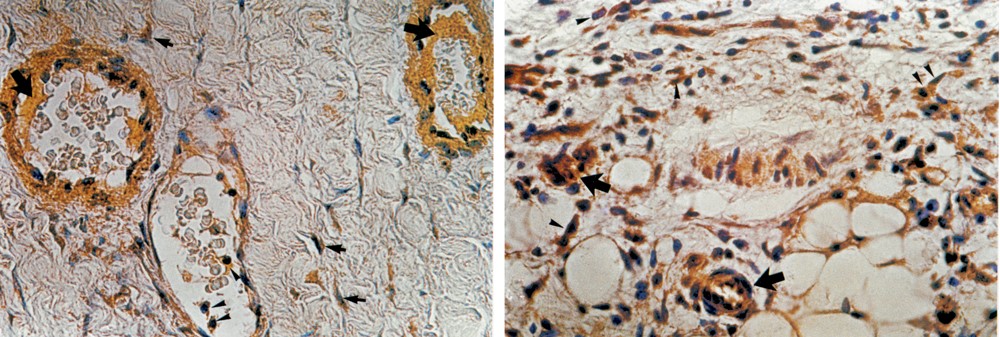 Figure 4a left: A borderline serous tumor. FasL-positive (brown staining)
endothelial cells, and muscle cells in the wall of small arteries and vein.
Figure 4b right: A mucous carcinoma as a metastasis in the great omentum. Fas + endothelial and muscle cells in the small arteries and fibroblasts. (Ref 77).
The role of immune reaction in cancer development and suppression
(Ref: 49 52 56 57 69 79-83 85 90-94 102 103 105 107 109 131 134 135).
We studied some cytological aspects of rat mammary cancer therapy with soluble tumor-associated antigens (sTAA- p66 and p51) and the anticancer drug cyclophosphamide (CPA). Vaccination with sTAA results in a significant increase in the production of CD8+ lymphocytes inside the tumors and in bone
marrow, and of CD8+ thymocytes in the medulla of the thymus. Treatment of rats with CPA sharply decreases the activity of lymph cells in tumors, especially of CD4+ lymphocytes. In the bone marrow, CPA affects the process of myelogenesis and causes significant substitution of cellular components with fatty tissue. The combined treatment with CPA and sTAA increases the number of lymph cells and the apoptotic index in tumors, and restored the rate of B cells producing in the spleen and in lymph nodes. In the thymus, CPA alone or in combination with sTAA repairs the inhibitor effect of a carcinogen on synthesis of CD4+ and CD8+ thymocytes.
The Secretory Immune System
(Ref: 40 86 88 95 98 99 100 110 136 137 138) .
We have described at first in the literature the secretory (mucosal) immune system (SIS) in the human embryos and fetuses. In the fetal placenta, the secretory immune components (secretory component-SC, joining (J) chain, IGA, IgG and IgM) are located mainly in the syncytiotrophoblast and cytotrophoblast. The villous stroma containes different lymphocytes and many macrophages, many of them are IL-2 positive. In the maternal placenta, the secretory immune components are located in the decidual cells. We found that the placenta has two different secretory immune systems, one fetal and the other maternal. They differ in their structure and orientation, and represent the largest extracorporal immune system. The maternal SIS protects the mother against paternal antigens from the fetus, while the fetal SIS protects the fetus against maternal antigens and infectious agents entering from the mother.
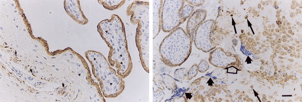
Figure 5 a left: Fetal and maternal SIS. Placenta of a 13- week-old fetus with low antigenic effect note the fetal SIS. Fifure 5 b right: Placenta of 17-week-old fetus with moderate antigenic effect IgA in maternal SIS (Ref 84).

Figure 6: Positive J chain immunostaining in the mucosal epithelium and in the endothelium of capillaries of the submucosal layers. (Ref 137).
Research collaborations with Weizmann Institute of Science
(Ref: 9 12 14 42 48 51 58-60 63 65 104 106 108).
Hematopoietic stem cells reside in the bone marrow where they proliferate and differentiate to replenish mature blood cells through the entire adult life. Stem cells are clinically used to repopulate the bone marrow of leukemic patients. For this purpose, stem cells are mobilized
from the bone marrow of healthy donors, and home to the bone marrow of the patients, to
repopulate it with multilineage blood cells.
The scope of our collaboration is to reveal mechanisms that regulate in vivo homing and
mobilization of hematopoietic stem cells, using the high repopulating potential of stem cells
isolated from cord blood and the immune deficient NOD/SCID mice, serving as a
preclinical small animal model.
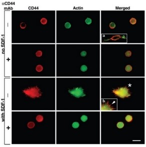
Figure 7: CD44 is localized to the edge of SDF-1- induced cell protrusions in HPCs
adhered to HA. The arrow is pointing to the intense staining of CD44 AT the edge of an SDF-1-induced protrusion (Ref 104).
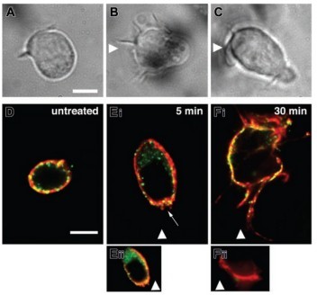
Figure 8: CD44 is localized to the leading edge of polarized human HPCs migrating toward SDF-1 (Ref 104).
Collaborative works were conducted in recent years with the other medical centers and groups as shown above.
• Prof. Igor Zusman, Koret School of Veterinary Medicine, Hebrew University;
• Prof. A. Ornoy, Department of Anatomy, Hebrew University Medical School;
• Prof. Y. Burstein, Organic Chemistry, Dr. F. Kohen, Biological Regulation Research,
• Prof. T. Lapidot, Immunology, Prof. Y. Reisner, Immunology, Weizmann Institute;
• Dr. Tendler, Clinical Chemistry, Rambam Hospital;
• Prof. S. Lundgren, Department of Oncology, University Hospital of Trondhum, Norway;
• Dr. J. Phipps, Nuneaton Hospital, Nuneaton, UK;
• Prof. F. Naftolin and Prof. G. Mor, Yale University School of Medicine, New-Haven, USA;
LEM (laboratory of early detection)
I have established and have managed the Laboratory for Early Detection of gynaecological cancer, since 1990.
Some interns conduct their thesis research in the laboratory under my guidance and supervision. Some residents conduct their basic science under my guidance.
